

+91 - 9818704499
+91 - 9311444806
+91 - 9311444806
Even now, after all these years, she can realize the moment when she first spotted a numb skin patch on her back. That was a coin sized white spot that had no feelings. This was an early phase of a disease called melanocyte.
Melanocyte, commonly known as vitiligo is a type of skin disease that affects a 2% of human population in India, every year. Though, this is not contagious it needs proper treatment to help control further spread of illness. Those, who are in the early phase of melanocyte or having their near and dear ones struggling with the disease may want to know what exactly melanocyte is. Let’s dig deeper in to the matter.
Definition: Cells of Neural crest origin found predominantly in the basal cell layer of the skin that helps in synthesis of melanin from tyrosine.
Mechanism of action
The melanin pigment is packaged in the melanosomes and these are transferred to the surrounding keratinocytes through the dendritic processes.
Ephelis
Ephelis is commonly known as Freckles and a common yet harmless lesion seen among the whites. These are red or light brown hyper pigmentation of the skin resulting due to the increased amount of basal keratinocytes and melanocytes.
Lentigo
Benign hyperplasia of the melanocytes occurring at all age but mostly seen in children and with no specific predilection of sex. This lesion includes oval, tan brown macules that appear like freckles but do not darken when exposed to light.
Melasma
These are mask like area of hyper pigmentation commonly seen in pregnancy.
Clinical appearance of areas of poorly defined blotchy macules in the cheeks, temples and forehead regions bilaterally is the main signs. Oral contraceptives, Hydantoin administration are some of the other etiological explanation to Melasma
Vitiligo
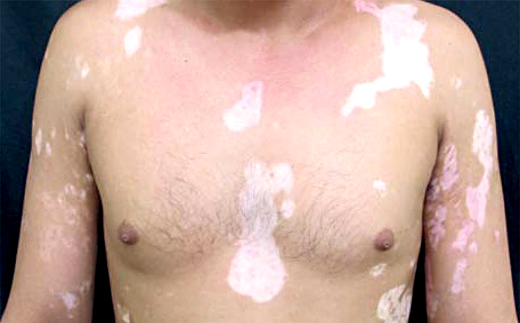
This disorder occurs as a result of partial or complete loss of melanocytes. It is also known as Lukoderma. It can occur unilaterally as well as bilaterally
Vitiligo can cause minor to extensive changes in the skin. Rate of occurrence is about 3 percent to 4 percent of the entire population. Hereditary pre disposition is common in over 30 percent of the sufferers with the presence of first sign by the age of 20.
Vitiligo consists of white symmetrical areas with darkened distinct borders. Hands face and areas that folds like armpits and elbows are affected most. Other most common sites are lips, orbits, back of the hands, nipples etc. besides the sun exposed sections.
Clinically the lesions are flat and well demarcated zones of pigment loss known as macules. The area of lesion may remain between few centimeters to over 100 centimeters. Wrists, Axillae, Peri oral and Peri orbital region are most affected. The progress is very abrupt in nature and may stop randomly after years of detoriation or vice versa.
Jennie..., whom we meet in the first paragraph of this write up, is still wondering why she had had vitiligo. Let’s find answer to her query.
Pretty well aware of her illness, now, Jennie decided to go for treatment to get rid of hideous white patches on her skin that make her look ugly.
Vitiligo treatment is believed to be the best in India at Delhi. Dermatologists carry out intricate procedures to provide apt possible perfection. Proper psychological counseling is carried out to guarantee mental stability after the treatment.
The numerous approaches for handling Vitiligo treatment in Delhi are:
Some of the age old techniques that is believed to give a small percentage of success includes Healing derma abrasion, laser ablation, and confined application of phenol or tri-chloro acetic acid.
Diagnostic investigations of Melanocytic Disorders
Histological finding specific to melanocytic lesions
Management
Prevention
 Increasing number of men and women who want to get rid of droopy upper eyelids and lower lid bags are achieving rejuvenated and young they desire from a special cosmetic surgery
 We, a team from Care well Medical Centre have been in Hair Transplantation for last 10 years. Initially FUT or Strip method was used. From last 4 years FUE is gaining popularity.
 Care Well Medical Centre is a renowned cosmetic surgery, plastic surgery and hair treatment center in South Delhi in CR Park touching nearby areas
 Carboxy therapy mentioned to the administration of carbon dioxide gas in cutaneous and subcutaneous nerves for therapeutic purposes.
 A fat grafting process involves transfers of fat from excess fat areas i.e. outer thighs in and injects it into areas that may be lacking in volume i.e. face, hands, breasts or buttocks.
 Cavitation is body sculpting with out anesthesia, with out scars, with out discomfort, with out down-time and presents a risk-free substitute to liposuction.
 CARE WELL MEDICAl CENTRE conducts Camp in Mandakani Welfare association, Chitranjan Park on 16th August 2014. The camp was headed by Dr. Sandeep Bhasin and his team.
 Even now, after all these years, she can realize the moment when she first spotted a numb skin patch on her back.
 Address: Care Well Medical Centre, 1, NRI Complex, Chittaranjan Park, New Delhi - 110019
Phone: +91-9818704499, +91-9311444806
Email: info@carewellmedicalcentre.in
Copyright © 2014, Carewell Medical Centre. All Rights Reserved
